WHY ROOT CANAL THERAPY?
The goals of root canal therapy are to:
- Remove bacteria and infected pulp form the pulp chamber and root canals
- Completely fill the canal(s) and pulp chamber with a solid filling material to prevent future trouble
When root canal therapy is done, inflammation in the bone around the root ends heal, and the tooth is saved. If you’re interested in a root canal procedure, please schedule your consultation today at our office in Tampa, Florida.
- Anatomy
- The Pulp
- Pulp And Jaw Infection
- Infection of the Face and Neck
- Why Root Canal Therapy?
- How Root Canal Therapy is Done
- A.Preparing the Root Canal
- B.Filling the Canals
- Restoring the Tooth After Root Canal Therapy
- Placing Posts
- Common Questions and Their Answers
- Accessory Root Canals
- Badly Curved Roots
- Very Narrow Canals
- Cracked Root
- Conclusion
Anatomy
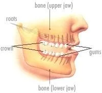
Each tooth consists of two parts: the crown and the roots. Only the crown is visible in the mouth. The roots are in the bone, under the gums. The gums are a protective type of skin that clings to the necks of the teeth and covers the bone holding the teeth. Molars are back teeth. They usually have two or three roots. Most other teeth have one root.
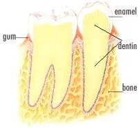
The crown of a tooth has an outer shell made of a very hard substance called enamel. The inside of the tooth is made of a less hard substance called dentin.
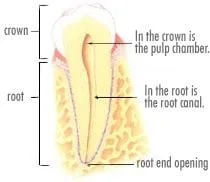
The center of the tooth is hollow. This hollow in the crown portion of the tooth is called pulp chamber. In the root portion of the tooth, the hollow narrow to become a small canal called the root canal.
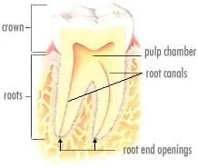
In teeth with more than one root, each root has its own canal that extends from the singe pulp chamber. Each root canal ends at a tiny opening at the end of the root.
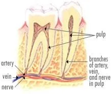
Usually there is only one canal in each root, but some teeth have more than one.
back to top
The Pulp
back to top
The Pulp and Jaw Infection
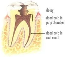
Bacteria are the most common causes of inflammation and infection of the pulp. They enter the pulp through tooth decay or if a tooth breaks.
Invading bacteria first overwhelm the pulp defenses in the pulp chamber. Then they destroy the pulp in the root canals.
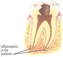
Toxins (poisons) from the bacteria that have destroyed the pulp can leak out of the root ends into the jawbone. The jawbone, like all bones, is a living tissue. It has arteries, veins, and nerves, like any other tissue. Therefore, it can become inflamed and infected by the presence of bacteria and their toxins.
back to top
Infection of the Face and Neck
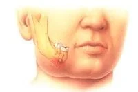
Finally, long-standing dental infection in bone can erode through the side of the bone into the mouth, or into the face or neck, to cause sudden, serious, and painful swelling.
back to top
Why Root Canal Therapy?
The goals of root canal therapy are to:
1. Remove bacteria and infected pulp from the pulp chamber and root canals
2. Completely fill the canal(s) and pulp chamber with a solid filling material to prevent future trouble
When root canal therapy is done, inflammation in the bone around the root ends can heal, and the tooth is saved.
back to top
How Root Canal Therapy is Done
The method of root canal therapy shown here is a very common one. Other techniques differ in some details and material, but accomplish the same goals.
Root canal therapy proceeds in two stages :
- Preparing the root canal
- Filling the canal
back to top
A.Preparing the Root Canal
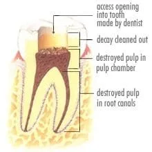
Step 1: Opening the tooth
The dentist gently makes an opening into the tooth. Local anesthesia (Novocain®) may be necessary to prevent root canal pain that can occur if any nerve fibers are still alive in the pulp.
All tooth decay is removed.

The dentist uses a series of very delicate, flexible finger-held or motor driven instruments.
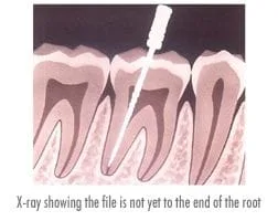
The one used in the illustration is a file.
Each file the dentist uses is slightly larger than the preceding one. The canals are delicately cleaned with these instruments to remove dead pulp debris and bacteria.
The dentist then shapes each canal to receive a filling.
X-rays help assure that the instruments go exactly to the end of the root and not beyond.
Canal preparation may take several visits, especially for difficult curved or narrow canals.
After preparation, all canals must be solidly filled. Otherwise, tissue fluid from the bone could eventually seep into any unfilled areas of the canal, decaying there into toxic products. These toxic products will then seep out of the root end into the bone to cause more inflammation.
back to top
B.Filling the Canals

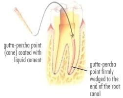
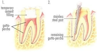
Step 1:The first gutta-percha point is inserted into the prepared canal. It matches the size of the last and largest file used.
Step 2: The dentist coats this point with a special liquid cement. The coated point is then inserted firmly to the end of the root. Wedged tightly, it completely seals off the end of the canal so that no fluids can leak past it.
Step 3: The dentist now packs the remaining portion of each canal with gutta-percha pieces up to the level of the pulp chamber.
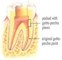
Step 4: Lastly, the dentist fills the tooth with a temporary protective cement.
back to top
Restoring the Tooth After Root Canal Therapy
Now that root canal therapy is finished, the dentist can repair the broken-down tooth crown that was damaged by decay.
Tooth decay that was bad enough to let bacteria into the pulp usually has destroyed much of the crown. Cleaning and shaping the canals further weakens the tooth. Such a tooth may break during chewing unless repair includes an internal post support followed by a fully covering crown.
back to top
Placing Posts
There are many internal post placement methods, all requiring great care and precision. One approach for a badly broken-down lower molar is illustrated below.
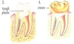
Root canal therapy is completed.
The temporary filling is taken out, and two-thirds of the gutta-percha is removed from the left root. A stainless steel or Titanium post is inserted.
At Agoka Dental, We almost always use Titanium Posts
A crown is precision fitted.
back to top
Common Questions and Their Answers
Q1. Is root canal therapy painful?
A1. Local anesthesia (Novocain®) can make most teeth painless to treat. Between treatments, aspirin-strength medications usually work well. Occasionally, a dentist at Agoka Dental will write a prescription..
Q2. Isn't root canal therapy quite expensive ?
A2. It's not, considering the time, patience, and skill needed to perform it. And the root canal cost is substantially less than the cost of a bridge needed to replace a tooth lost because root canal therapy was not done.
Q3. What is an endodontist ?
A3. An endodontist is a root canal therapy specialist. After four year of dental school, he or she takes two years of intensive specialty training. An endodontist does both routine and complicated root canal cases.
Q4. Despite a dentist's or endodontist's best effort , don't some root canal therapies fail ?
A4. Seldom. The odds for success in uncomplicated cases are excellent; in fact, they are well over 90 %.
Q5. Why do the rare failures happen ?
A5. Usually due to special complications. Illustrations of some these complications follow.
back to top
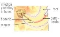
Accessory Root Canals
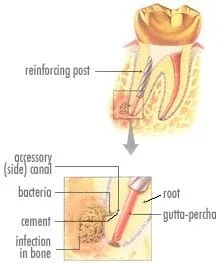
Some teeth have large side canals coming off the main canal, most often near the root end. These cannot be cleaned out, and the bacteria they contain may keep a root-end inflammation going after the main canal is filled.
back to top
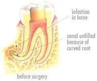
Badly Curved Roots
back to top
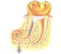
Very Narrow Canals
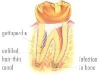
Some canals become exceedingly narrow, especially in the teeth of older people, or in teeth with deep filling.
It was impossible to prepare the tiny lower ends of the canals of this tooth. These canals, each only the width of a hair, contain bacteria that can cause continuing trouble.
back to top
Cracked Root
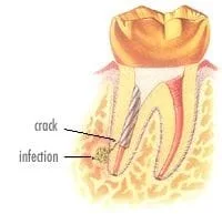
A tooth can develop a hairline crack in its root, a crack so small it won't show on an X-ray, but still large enough to harbor bacteria that can do damage.
Q. Does failed root canal therapy mean the tooth must be extracted (taken out)?
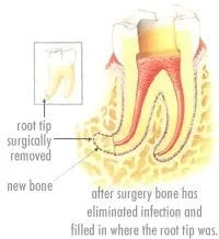
A. Sometimes Yes, but Usually not. There are special techniques... of canal retreatment. And, when a good seal at the end of a root is impossible to attain, the root end can frequently be surgically removed to solve the problem.
Usually the treating Dentist and/or an Endodontist will evaluate individual cases and decide on the course of action at this point.
Conclusion
Root canal therapy saves teeth with infected pulps. It avoids the complication and greater expense of replacing teeth that would otherwise be lost.
back to top